Invitrocue’s expertise in 3D culture allows us to successfully recapitulate in vitro the normal and disease conditions of humans. Our 3D cell culture techniques using our 3D Cellulose Sponge scaffold, or non-scaffold-based techniques are more cost-effective and physiologically relevant compared to conventional 2D monolayer cultures.
By co-culturing and tri-culturing organ-specific cells with immune cells or endothelial cells, our 3D models are better able to recapitulate the microenvironment and functions of human organs, providing a more clinically relevant model for drug efficacy and toxicity testing.
Our 3D Cell Culture Platform
2D cell culture
Lack of cell-to-cell and cell-ECM interaction
No gradients present
No drug resistance
Co-culture unable to establish a proper microenvironment
Poor clinical correlation
3D cell culture
Physiologic cell-to cell and ECM interaction
Drugs, oxygen, and nutrients diffuse in gradient
Drug resistance in vivo
Co-culture of multiple cell types mimic in vivo microenvironment
More reliable and better estimate of in vivo responses
Respiratory models
Invitrocue offersin vitro 3D respiratory models using air-liquid interphase culture for drug testing, inhalation toxicity, viral infections and inflammation studies.
Features:
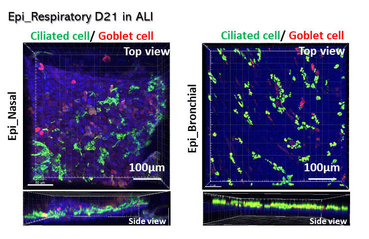
Our 3D mucociliary tissue model consists of normal, human-derived nasal/bronchial epithelial cells
Our3D structure consists of basal cells, goblet cells and ciliated cells with mucociliary clearance functions
Ourmodel exhibits pseudostratified columnar epithelial morphology with high uniformity and reproducibility
Our culture system preserves the physiological characteristics of nasal and bronchial epithelia
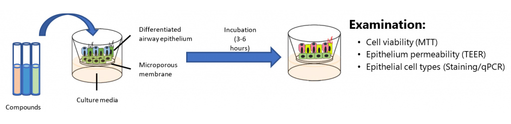
Inhalation Toxicity Test
Our inhalation toxicity test evaluates the toxic characteristics of inhalable materials, such as gases, volatile substances or aerosols/particulates. Acute inhalation toxicity data are used to satisfy hazard classification and labelling requirements, to estimate the toxicity of mixtures, and to assess human health and environmental risks.
Experimental design for drug testing using nasal/bronchial models
Test Model
Nasal/Bronchial models
Replicates
N=3 tissues per test condition
Exposure Time
3-6 hours topical exposure to a predetermined 4-concentration range of test chemicals
Assay Controls
Negative Control – Sterile DI H2O
Positive Control – 14mg/ml Formaldehyde
Endpoints
MTT, TEER, Marker staining, real-time PCR
Data Delivery
Cell viability, permeability analysis,cell type and gene analysis
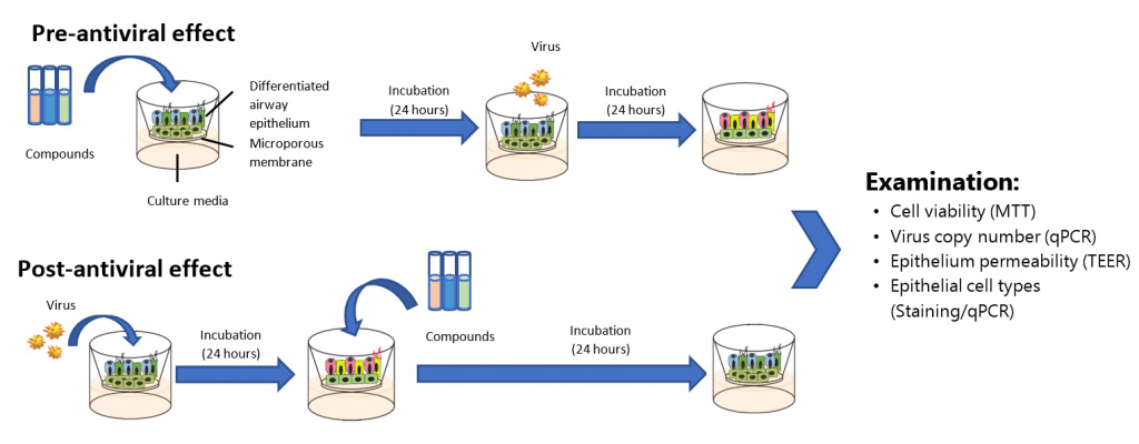
Anti-Viral Test
Our viral infected model for common cold (Rhinovirus) and flu (influenza virus) can help to evaluate the efficacy of antiviral drugs in inhibiting viralreplication, enhance mucociliary clearance, and elevate the innate immune defence of airway epithelium.
Confocal image of 3D nasal and bronchial models
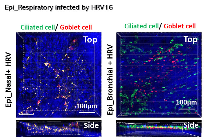
Confocal image of infected 3D nasal and bronchial models. Decrease in ciliated cells and increase in goblet cells observed.
Experimental design for drug testing using viral-infected nasal/bronchial models
Test Model
Nasal/Bronchial models
Replicates
N=3 tissues per test condition
Exposure Time
24-hour topical exposure to a predetermined 4-concentration range of test chemicals
24-hour exposure to virus
Assay Controls
Negative Control – Sterile DI H2O
Positive Control – MOI 10
Endpoints
MTT, TEER, Marker staining, real-time PCR
Data Delivery
Cell viability, permeability analysis,cell type and gene analysis
Wound healing and Inflammation models
The wound healing process involves the repairing of skin homeostasis. It is a complicated process involving multiple interlinked phases such as haemostasis, inflammation, proliferation, and remodelling which generally takes around 4 weeks.
Obstacles in wound healing can occur from internal factors such as diabetes, coronary artery disease, aging, stress and immune system related diseases, as well as external factors such as bacterial infections, medication, smoking and nutrition. If not treated properly, these factors may lead to chronic non-healing wounds and resulting complications such as infection and tissue necrosis.
Invitrocue provides in vitro 3D skin models for the preliminary efficacy and safety evaluation for wound dressings, cosmetic products and skincare products.
In vitro 3D wound healing models
Our in vitro 3D skin models are composed of normal human epidermal keratinocytes (NHEK) and normal human dermal fibroblasts (NHDF)—cell types that mimic the wound bed. Interaction of keratinocytes and fibroblasts is important during the rebuilding of tissue integrity. Fibroblasts play an important role in wound healing by supporting the expansion and migration of keratinocytes.
Our 3D skin models are superior to the conventional monolayer model as they better recapitulate in vivo epidermis and skin barrier functions.
Advantages of our model:
More complex model than the 2D scratch wound assay
Cells are grown in a 3D collagen matrix that can provide structural support for the skin model
Allows for the study of cell migration toward the wound bed with the use of 2 major cell types (NHEK and NHDF)
Flexible model that can induce inflammation and mimic diabetic wound conditions by modifying the composition of the culture medium
Applications:
Wound dressing
Drug evaluation
Cosmetic products
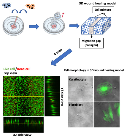
The diagram above shows the method of wound preparation (gap creation) in our 3D co-culture of keratinocytes and fibroblasts. The wound healing and cell migration can be easily quantified through the measurement of gap closure over the time.
Reference: Jonkman, James E N et al. “An introduction to the wound healing assay using live-cell microscopy.” Cell adhesion & migration vol. 8,5 (2014): 440-51. doi:10.4161/cam.36224